Sometimes the transition switch from conventional microscopy to digital pathology is made challenging for the pathologist by differences in what the viewed images look like over the two mediums. So, what do you do to make a great digital microscope image?
Sometimes the transition switch from conventional microscopy to digital pathology is made challenging for the pathologist by differences in what the viewed images look like over the two mediums. So, what do you do to make a great digital microscope image?
No amount of processing can make up for a less than great raw image, the material must be of very high quality. A high-quality imaging system with a well-integrated objective lens and components make a great starting point, but what really makes a difference is how the number of adjacent photographs of the sample on the glass slide are captured and processed.
What makes the Grundium Ocus® microscope slide scanner special is that it doesn’t just filter colors from a regular image, but instead red, green, and blue are all photographed individually in full resolution. This ensures a far superior, true color reproduction instead of the common approximation. Different staining techniques appear natural as if viewed on a microscope and make for easy collaboration and analysis of scans between experts.
If you’re capturing images of samples with varying thicknesses, such as tissue, at a constant distance, some parts of the sample may be out of focus. To mitigate this, the Ocus has electronic autofocus. As it captures images, it is constantly following the sharpness, predicting the elevation from the adjacent squares. If the image isn’t sharp, the Ocus automatically expands the focus search range to return to focus. The focus is built in a dynamic and adaptive way, which adapts to slide tilt and sample topography, adding to the clarity of the scanned image.
The Ocus has an artificial intelligence algorithm, which detects potentially interesting areas in the overview image. When the user selects their area of interest, the Ocus uses the detected sample regions in the overview image to guide scanning and focusing. This is yet another way the Ocus works smartly to make the work of the pathologist faster, easier, and more convenient.
A microscope scanner basically divides the scanning area into a grid, and then takes a high-resolution image of each part. A viewable image is then stitched together from those individual photos. Because there is always a minute overlap at the seams between photos, they are “stitched” together digitally. If each photo is matched to the next linearly, it can result in inaccuracies, with rows of photos skewing off and not matching at their ends. The Ocus uses a patented Grundium stitching model known as Global Optimization, where every photo is matched to all other photos in the entire scan area, not just the previous. This results in a highly accurate, undistorted image like no other!
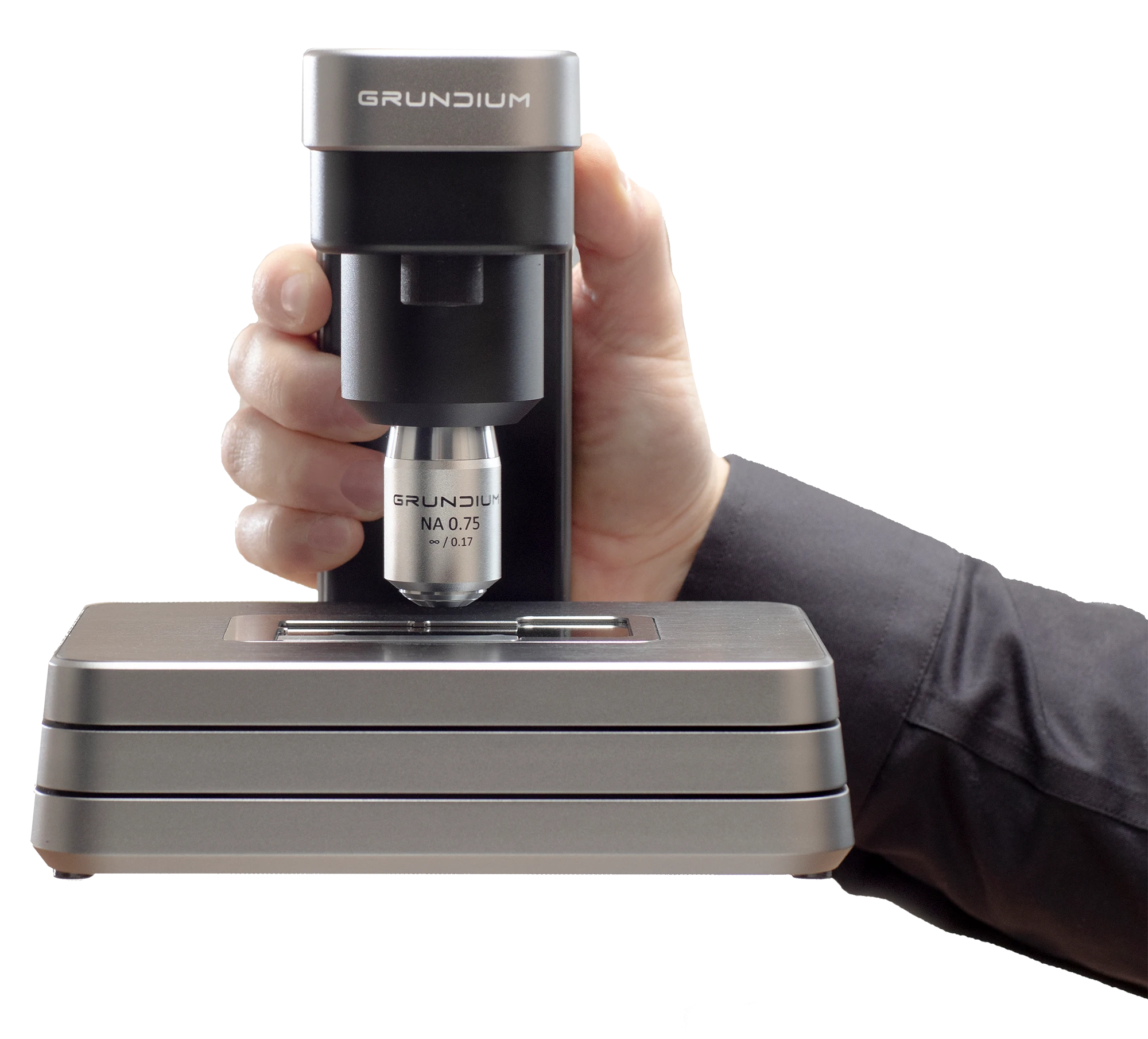
Each Grundium Ocus® scanner is tested to the highest standard before it is shipped to a customer. In calibration, each scanner is adjusted for variations in lens distortion, shading, focus correction, image magnification, stage movement, camera alignment, and LED luminosity, and adjusted accordingly. This ensures every single scanner leaves the assembly line up to a tight specification.
The Grundium team has its roots in Nokia mobile devices. This background gives us a unique edge in designing beautiful, robust, compact, and practical high-precision optical instruments and manufacturing them to a rigorous quality specification. Grundium employs experts in many different disciplines. In addition to all the know-how that goes into manufacturing the devices, especially important for making state-of-the-art digital scanners are optics, signal processing, and mathematical optimization. All this expertise combined enables us to look outside the box and create solutions that are smaller, cleverer, and more practical than anything else.
The Grundium Ocus® is the most practical microscope scanner in the world. Its small footprint means it can find a spot in any lab. The Ocus® is so simple to use anybody can be trained to operate it in just 15 minutes. Created by ex-Nokia flagship camera phone engineers, it takes the sharpest images in the business. It is much more affordable than big multislide scanners. Its software is so advanced it can be integrated with any existing LIS and it supports the most common file formats. The Ocus scanners simply remove the entry barrier to digital pathology.
If you are looking for a small footprint and sharp imaging component for your pathology solution or if you want to hear more about how to integrate an Ocus scanner into your system, please contact us.