Ocus®40: High magnification with utmost precision

Divider
The Ocus®40 microscope slide scanner delivers high-resolution imaging with powerful 40× magnification, offering a high-performance solution for cytology, histology, and detail-critical applications.

Designed for demanding workflows, it combines precision, clarity, and ease of use.
  • Magnification: 40x
  • Resolution: 0.25 µm/pixel
  • Capacity: 1 standard slide (1″ × 3″)
  • Automatic tissue detection
  • Continuous autofocus
  • Z-stacking up to 5 layers
The Ocus®40 digital pathology slide scanner designed for digital pathology workflows, delivering high resolution imaging with exceptional detail.

A versatile tool suitable for a variety of pathology APPLICATIONS

The Ocus®40 microscope scanner is a versatile tool designed to enhance digital pathology workflows with high-resolution imaging. It supports a wide range of applications, including:

  • Histopathological diagnosis
  • Tumor grading and staging
  • Immunohistochemistry (IHC)
  • Biopsies and Fine Needle Aspirations
  • Cytology

With its advanced features, the Ocus®40 ensures accurate and efficient evaluation of tissue samples, making it an essential asset for modern pathology labs.

Designed for both accessibility and high performance, the Ocus®40 combines quality, affordability, and user-friendliness. This level of magnification unlocks new possibilities in digital pathology, allowing for in-depth analysis and interpretation of samples with unparalleled clarity.

NEW; Ocus®40 Sample slide gallery

Explore our new image gallery featuring sample slides scanned with the Ocus®40. View high-quality images in an online viewer or download SVS files for further analysis.

Unmatched Ease of Use

Designed with the user in mind, the Ocus® simplifies the complex. Its intuitive, browser-based interface and streamlined workflow allow for quick scanning, viewing, and sharing of slides, making it accessible to users of all skill levels. From setup to operation, the Ocus® is designed to remove barriers, enabling professionals to focus on their critical work without the hassle of complicated equipment. This ease of use is a cornerstone of the Ocus® design, reflecting Grundium's philosophy of making powerful technology accessible to everyone.

Intuitive Design

Jump straight into your work without the hassle. The Ocus' user-friendly, browser-based interface and streamlined operations allow you to scan, view, and share slides with just a few clicks.

Small footprint

The compact size – just 7 inches in all dimensions – allows The Ocus to comfortably fit even on the most cluttered lab desk, eliminating the need for dedicated equipment space.

Rapid Setup

Go from unboxing to scanning in just 15 minutes. Spend less time setting up and more time on what matters most.

See the Ocus® in action

Curious about the Ocus® user interface and how easy it is to use? Look no further! Watch our 10-minute walkthrough video, where we showcase the key features of the device and its user-friendly interface. Check it out below!
We’d be happy to arrange a personal online demo for you! If you're interested, simply reach out to request a demo, and one of our experts will get in touch. The demo is free of charge and comes with no obligations.
Illustrative image representing the Ocus®40 as an affordable digital pathology solution that supports a streamlined workflow and efficient operation.

AN AFFORDABLE INVESTMENT THAT BRINGS Exceptional Value

The Ocus® scanners represents a significant advancement in cost-effective digital pathology. They are designed to provide exceptional value, offering premium features and performance at a fraction of the cost of traditional systems.

This affordability does not come at the expense of quality or functionality; instead, it opens up new possibilities for institutions and practices of all sizes. By minimizing the total cost of ownership, the Ocus® ensures that resources can be allocated efficiently, making advanced digital pathology accessible to a wider audience.

Designed for Diverse Applications

The Ocus®40 embodies the essence of versatility and precision in digital pathology, engineered to meet the demands of remote consultations, research, and education. Its groundbreaking 40x magnification brings forth an unprecedented level of detail and clarity, making it an invaluable tool across a wide spectrum of applications.

The Ocus®40 is more than just a microscope slide scanner; it is a multifaceted tool designed to support and enhance work in diagnostics, research, and education, reflecting Grundium’s commitment to innovation and excellence in digital pathology.
A

REMOTE SECOND OPINIONS AND CONSULTATIONS

The Ocus®40 bridges the gap between specialists, enabling the sharing and analysis of high-quality digital slides in real-time, thus facilitating second opinions and collaborative diagnoses without geographical constraints.
Ocus®40 for second opinions
University professor showing an Ocus scan result on a lecture

RESEARCH AND EDUCATION ENVIRONMENTS

The Ocus®40 enhances research and education by capturing high-resolution scans for detailed specimen analysis, advancing scientific knowledge, and transforming learning with digital slides that improve material quality and support remote learning, making specialized knowledge more accessible.
ocus®40 in Research & education applications

Send us a message!

Do you have a question about our devices? Wondering how the Ocus® scanners could be implemented in your workflow? Whatever it is, we are happy to hear from you!

Send us a message with the form here, and our experts will get back to you shortly!

If you need assistance with your device, please contact Grundium's customer support.

Why Choose The Ocus®40?

Choosing the Ocus®40 microscope slide scanner elevates your pathology practice with unmatched precision and efficiency. Designed for cutting-edge digital pathology, the Ocus®40 makes highly detailed diagnostic information accessible to professionals worldwide. Its advanced 40x magnification ensures superior clarity and depth, meeting the stringent demands of modern diagnostics.

Redefine your standards with the Ocus®40, where exceptional image quality and operational flexibility empower laboratories to achieve outstanding results in cytology, histology, and beyond.
The compact Ocus®40 slide scanner held on a user’s palm, highlighting its intuitive design and suitability for scanning glass slides in modern digital pathology workflows.

want to know more?

Send us a message and we
will get back to you soon.

Ocus®40 Technical specifications

Magnification: 40x

Numerical Aperture: 0.75

Resolution: 0.25 µm / pixel

Depth of field: 1 µm

Slide format: 75 x 25 mm (3 × 1 in)

Scan speed: ~200 sec / 15 × 15 mm

Image formats: .SVS, .TIFF, .SZI

Focusing: Fully automatic

Image sensor: 12 MPix

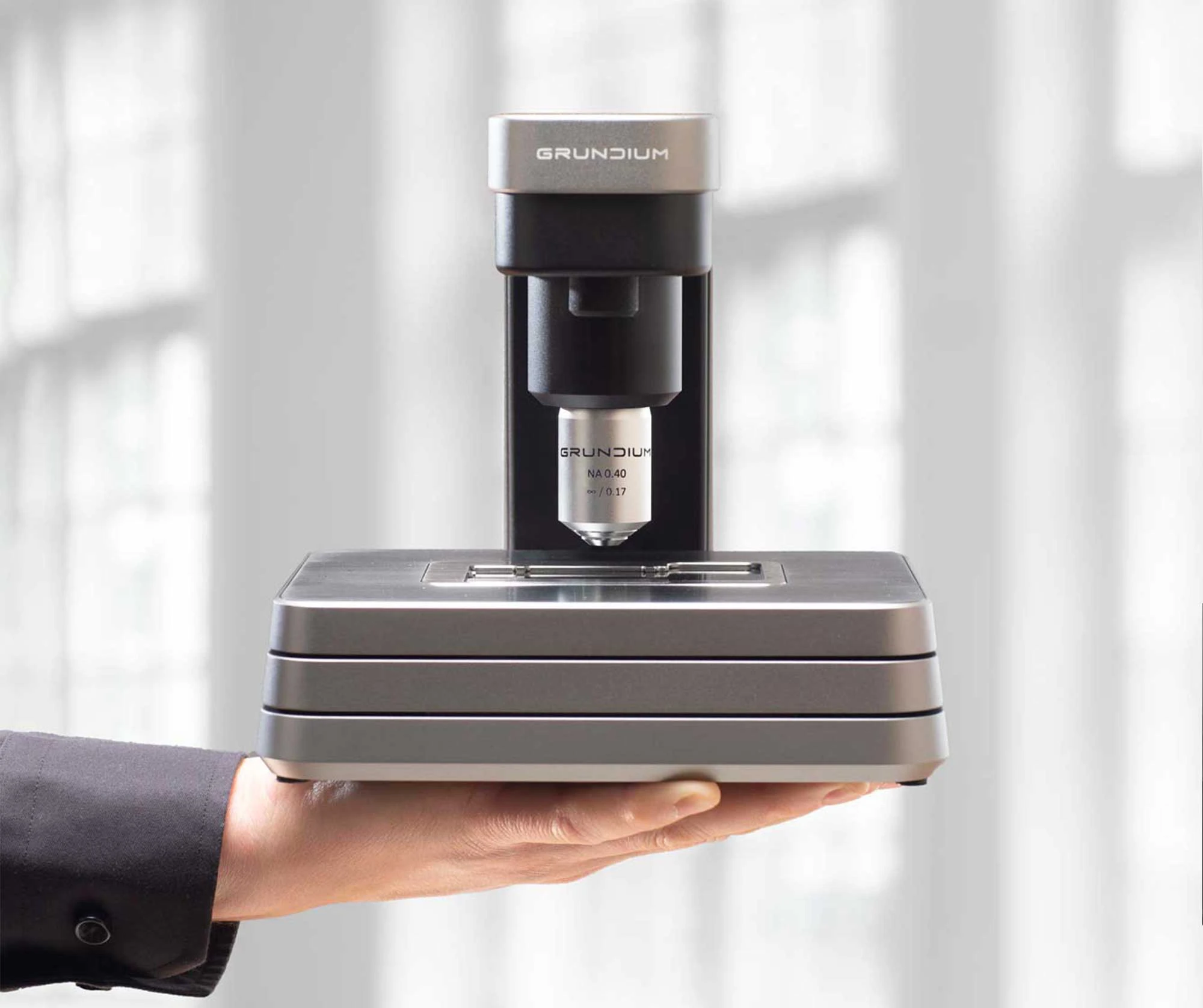
Dimensions: 7 × 7 × 7.5 inches (18 × 18 x 19 cm)

Weight: 7.7 Lbs / 3.5 Kg

Internal Storage: 500 Gigabytes

Power consumption / Standby: 0,03 A, 0,007 kVA, 0,007 kW

Power consumption: / Operation: 0,09 A, 0,021 kVA, 0,021 kW 

The Grundium Ocus® microscope slide scanners are CE-marked under the In Vitro Diagnostic Regulation (IVDR, EU 2017/746) in the European Union, making them approved for use in clinical and diagnostic workflows within the EU. In the United States, the Ocus® devices are currently classified as Research Use Only (RUO).

What our customers say

Dr. Alba Torres
Out of all the scanners I’ve used — and I’ve used a lot — it’s the easiest one. We've scanned more than 30 000 slides without any technical issues.
Dr. Alba Torres
Process and Quality Director
Cepatho, Brazil
Teppo Haapaniemi, Medical Cell Biologist, Fimlab
With Grundium Ocus-enabled telepathology, we can provide better service to our customers; the hospitals. For the hospitals the process doesn’t change, but they benefit from Fimlab working faster and more accurately with digital pathology.
Teppo Haapaniemi
Medical Cell Biologist, Pathology
Fimlab
Ben Cahoon, CEO, Techcyte
Not only has Grundium created something unique in the market with the beautifully designed small-footprint scanner, but they also have a super technical team that can work really close together to make the whole solution seamless and is willing to invest themselves in making something new. It’s super unique in the world to have everything from the region of interest, focus layers, depth of field and the whole experience totally integrated.
Ben Cahoon
CEO
Techcyte
Scott Rombach, Specialist Direct
The Ocus scanner is small and produces very sharp images fast. It is a vital instrument in field use in organ procurement. Its unparalleled portability helps save lives.
W. Scott Rombach
CEO
Specialist Direct Inc.
Julia Cara Thierauf, Research Fellow in Pathology
We have excellent images with the Ocus and I couldn’t be happier to have this little helper on my desk here. I use it mainly as a microscope, to be honest, but of course, scan my slides as well. It has really helped me so much.
Julia Cara Thierauf
Research Fellow in Pathology (EXT)
Massachusetts General Hospital
Fabio Papadia, Philips
The Philips IntelliSite Digital Pathology Solution has the capabilities to upload, view and annotate the images produced from the Grundium scanner. Zooming and panning is really fast, even when the images are physically stored on a server in the Netherlands and I’m viewing the images from a workstation in Sweden.
Fabio Papadia
Business Development Manager Nordics,
Digital and Computational Pathology
Philips

Want to learn how the Ocus scanners would ENHANCE your workflow?

Experience the future of digital pathology in a personalized 30-minute demo with a Grundium expert!

Tailored to your schedule, this session will help you discover the perfect Ocus® scanner for your needs, explore our pricing options, hear inspiring user success stories, and learn how an Ocus® could elevate your lab’s digital workflow.

Let’s transform the way you work – schedule your demo today!
Ocus®40 held in hand

Frequently Asked Questions – Ocus®40 Digital Pathology Slide Scanner

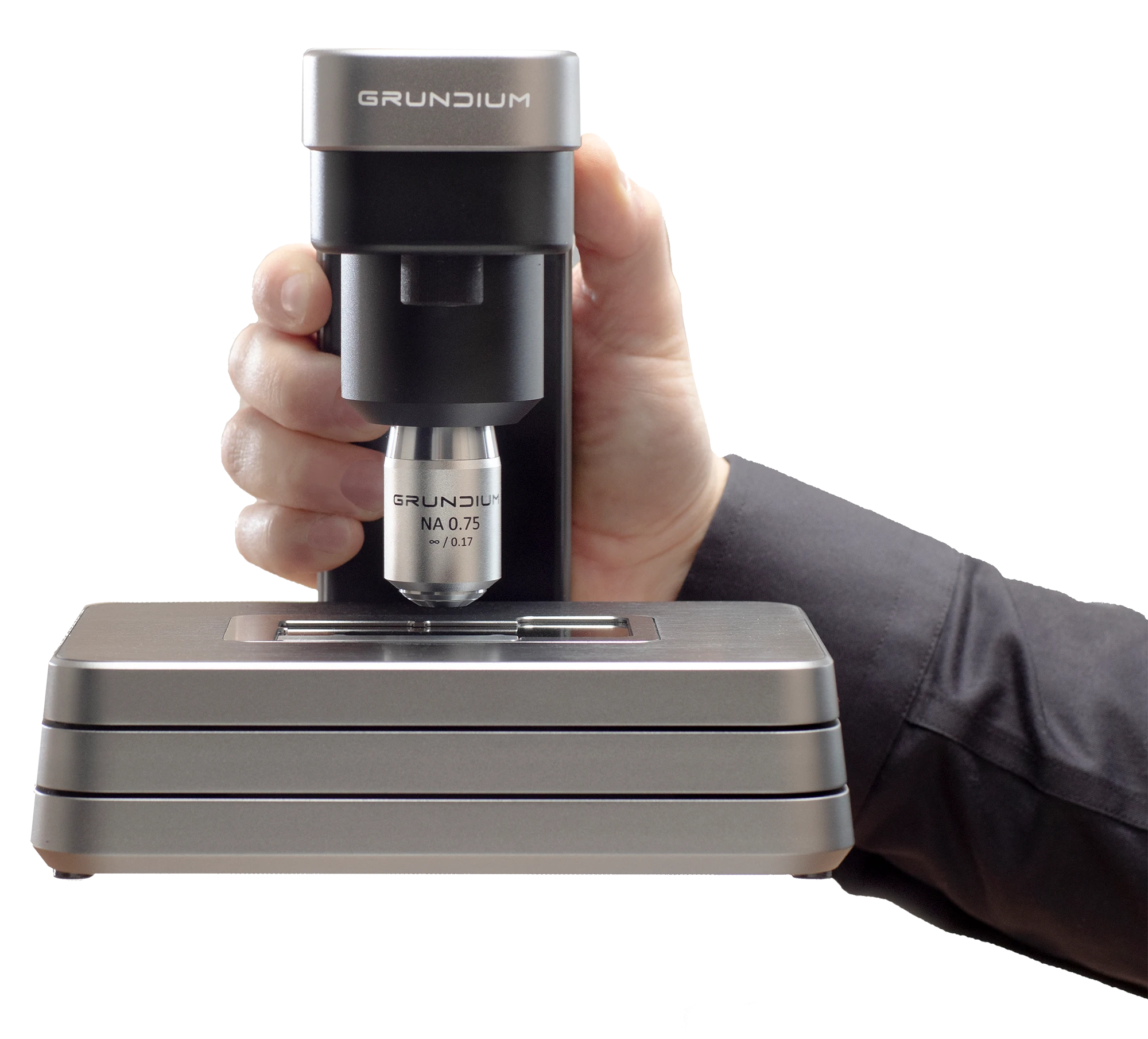
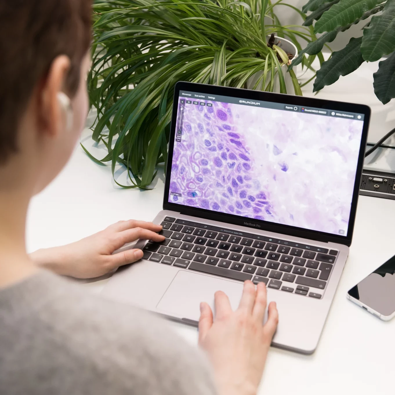
The Ocus®40 is a digital pathology slide scanner designed to digitize standard glass microscope slides at 40x magnification. It delivers high-resolution whole slide images that support diagnostics, research, and education, enabling precise digital visualization for applications that require exceptional detail.

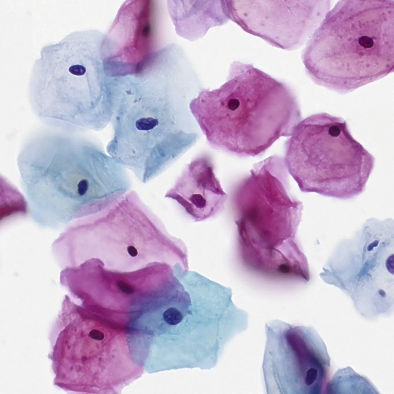
Ocus®40 is used to digitize glass slides for a wide range of digital pathology applications, including routine histology (H&E), immunohistochemistry (IHC), tumor grading and staging workflows, and biopsy review. Its higher magnification is also well suited for cytology applications such as fine needle aspiration (FNA) and other specimens where additional cellular detail is helpful. It is also used in research and education settings where high-quality digital slides support analysis, collaboration, and training.

The primary difference is optical performance and the level of detail captured. Ocus®40 scans at 40× (0.25 µm/pixel) with a higher numerical aperture (0.75) and ~1 µm depth of field, which supports closer evaluation of fine cellular detail. Ocus®20 scans at 20× (0.50 µm/pixel) with NA 0.40 and ~5 µm depth of field, which is commonly sufficient for routine histology review and provides a broader in-focus plane. 

In workflow terms, Ocus®20 is typically used when speed and data efficiency are priorities (about ~1 minute for a 15 × 15 mm area), while Ocus®40 is selected when maximum detail is needed (about ~3 minutes for the same area). Both are single-slide scanners for 1” × 3” slides, support automatic tissue detection, continuous autofocus, live view, and offer export in DICOM, SVS, TIFF, and SZI.

Yes. Ocus®40 is suitable for routine pathology scanning, particularly in workflows that benefit from higher magnification and detailed image quality. It can be used as a primary scanner for routine casework or as a complementary scanner alongside other systems, depending on the lab’s case mix, throughput targets, and standard operating practices.

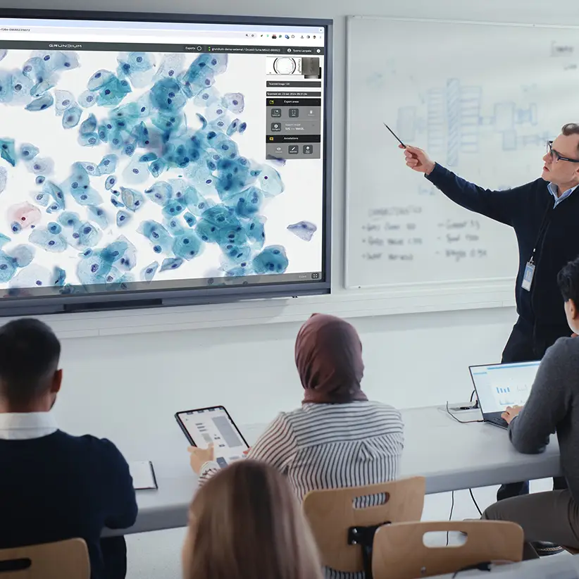
Ocus®40 enables remote consultations and second opinions by digitizing glass slides into high-quality whole slide images that can be shared for review beyond the originating lab. This supports collaboration between sites, access to subspecialty expertise, and multidisciplinary case discussions without the delays and risks of shipping physical slides.

Ocus®40 supports digital pathology workflows by providing streamlined slide scanning with consistent, high-resolution output in a compact, easy-to-deploy system. It integrates into routine laboratory work by enabling efficient digitization of cases and generating digital slides suitable for on-screen review, archiving, and downstream analysis.

OUR HEADQUARTERS

Grundium Oy
Saavutustenkatu 3
33720 Tampere
Finland
Ocus® products are designed
and manufactured in Finland 🇫🇮

Distributors

USA

Grundium scanners distributed by Fisher Healthcare in the U.S.

Brazil

Grundium scanners distributed by Fisher Healthcare in the U.S.

Turkey

Grundium scanners distributed by Fisher Healthcare in the U.S.

Korea

Grundium scanners distributed by Fisher Healthcare in the U.S.

INDIA

Grundium scanners distributed by Fisher Healthcare in the U.S.

Poland

Grundium scanners distributed by Fisher Healthcare in the U.S.

Greece

Grundium scanners distributed by Fisher Healthcare in the U.S.
Grundium earns the Future Workplaces 2025 certification
© Grundium Oy 2025. All rights reserved.