Whole slide imaging, also known as WSI, virtual microscopy or digital pathology, is a technique that involves capturing high-resolution digital images of an entire microscope slide. This technology revolutionizes the field of pathology by digitizing glass slides and allowing pathologists to view and analyze them on a computer screen.
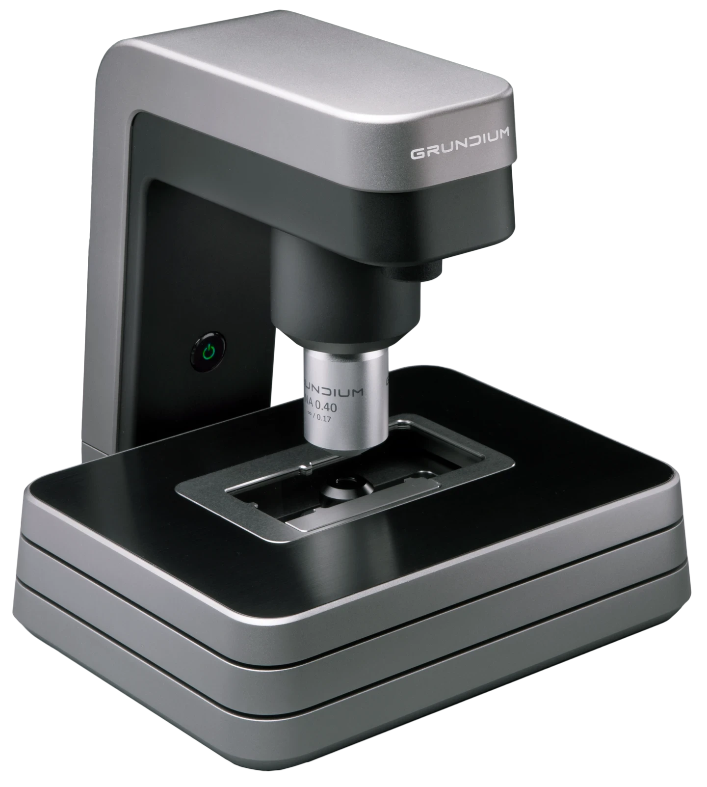
Slide scanners are the core devices used in whole slide imaging. They are designed to capture high-resolution digital images of entire glass slides. These scanners utilize advanced optics and digital cameras to scan the entire slide surface, capturing images of each field of view and stitching them together to form a complete, seamless digital representation of the slide. The resulting digital slide can be viewed, manipulated, and analyzed using dedicated software. Slide scanners vary in their scanning speed, resolution, and automation capabilities.
One of the key advantages of whole slide imaging is the ability to store and access large collections of slides in a digital format. This allows for easy retrieval of slides whenever needed. Digitized slides can be securely stored in digital archives, facilitating data management and retrieval for research, education, and archival purposes.
Whole slide imaging also enables remote viewing and collaboration. Pathologists can access digital slides from any location with an internet connection, allowing for telepathology consultations, second opinions, and collaboration among experts. This capability is particularly valuable for remote areas, where access to specialized expertise may be limited.
Additionally, whole slide imaging offers benefits such as enhanced image analysis and quantification, integration with computer-assisted diagnostic tools, and the potential for artificial intelligence-based algorithms to aid in automated analysis and pattern recognition.
Whole slide imaging is increasingly being adopted in pathology practices, research institutions, and healthcare systems worldwide, as it enhances efficiency, collaboration, and overall workflow in the field of pathology.
Want to say goodbye to the cumbersome process of mailing slides or waiting for courier services? With an Ocus® scanner at their fingertips, our customers instantly share high-resolution images with colleagues for consults, regardless of their geographical location.
The Ocus’ digital sharing capabilities make it easier than ever to collaborate on challenging cases, ensuring more accurate and timely diagnoses.
Our customers streamline their intraoperative consultations with Ocus® scanners, eliminating the need to travel between locations or wait near the operating room by having an Ocus® scanner conveniently located in a lab nearby.
You can view high-quality frozen section samples on your screen within minutes and effortlessly collaborate with colleagues, no matter their location. With Ocus®, our customers say they can focus on what truly matters — providing faster, more precise patient care — while saving valuable time for critical decision-making.