Artificial intelligence, AI, has been one of the most talked-about topics in pathology for years. AI stands on the shoulders of digital pathology, which can already feel threatening to the expert who has maybe spent decades looking at specimens in a microscope. Will all this rapidly developing technology make the expert obsolete?
As the demand for diagnostic tests surges—driven by an aging population and increasing cancer prevalence—pathology services are facing a challenge: a shortage of new pathologists. This gap in the workforce makes digital pathology not only a solution but a necessity in modern healthcare. The shift from traditional slide reviews to digital pathology is revolutionizing diagnostic services, offering faster, more accurate results while expanding access to expert opinions globally.
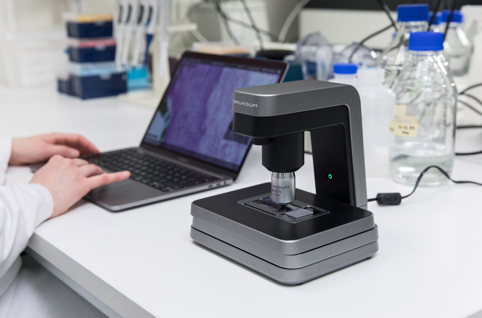
In conventional pathology, biopsies are processed and manually examined under microscopes, a labor-intensive and time-consuming method. With digital pathology, samples are digitized using high-precision microscope scanners, such as Grundium’s Ocus® scanners, enabling instant access to slides online. This digitization accelerates diagnoses and allows pathologists from different locations to collaborate more efficiently.
The introduction of artificial intelligence (AI) into digital pathology is transforming the way pathologists work. Machine vision systems are now able to pre-screen slides and highlight areas of concern, helping reduce human error and the time spent on routine tasks. AI-powered deep learning algorithms are designed to improve as they analyze more data, yet the integration of these systems into pathology is still in its early stages and requires careful calibration to achieve high accuracy.
However, AI in pathology is not about replacing human expertise—it’s about augmenting it. AI systems can handle repetitive and time-consuming tasks, freeing up pathologists to focus on more complex cases. This collaboration between AI and human pathologists ensures more consistent, accurate diagnoses while reducing the overall workload.
The combined power of digital pathology and AI offers several advantages:
Despite its potential, the transition to digital pathology faces several challenges. The implementation is costly and requires significant changes to existing infrastructures, including scanner integration and digital storage solutions. Furthermore, many healthcare systems are constrained by limited resources and regulatory frameworks that slow the adoption of digital tools.
The adoption of digital pathology, paired with AI, is poised to transform diagnostic services, providing pathologists with the tools to meet growing demands efficiently. While the path to full integration is complex, the potential benefits—including faster diagnoses, reduced human error, and enhanced collaboration—are undeniable. As technologies advance, digital pathology will become an essential element of modern healthcare, helping to deliver more accurate and timely diagnoses worldwide.
With the advancement in our understanding of diseases and the complexities of the human body, the demand for diagnostic tests has surged. This increase, coupled with a growing aging population facing a higher prevalence of cancer, has intensified the need for efficient diagnostic services. However, the field of pathology faces a notable challenge: a decline in the number of new pathologists entering the field. This global issue has strained the capacity to meet the rising diagnostic demands.
In traditional pathology, samples acquired during biopsies are sent to pathology labs, where they are processed and examined under microscopes by pathologists. This process can be time-consuming, depending on the proximity of pathology services and the nature of the case. Digital pathology offers a transformative solution by digitizing biopsy samples using high-precision microscope scanners. These digital images can be shared instantly online, enabling pathologists, irrespective of their location, to conduct diagnoses or seek second opinions efficiently. This technology not only accelerates the diagnostic process but also facilitates educational opportunities, allowing multiple pathologists to view and discuss the same sample simultaneously.
The transition to digital pathology opens avenues for integrating machine vision and artificial intelligence (AI) in diagnostic processes. Pathologists traditionally spend extensive time examining slides, a task that becomes increasingly demanding with higher workloads. Machine vision systems can assist by pre-screening slides and highlighting areas of interest, thus reducing the monotony and potential for human error.
When these systems employ deep learning algorithms, they evolve into more sophisticated AI tools capable of learning from vast datasets. However, the development of such AI applications is time-intensive and requires careful calibration to ensure accuracy. While AI holds great promise in pathology, it is not close to superseding human expertise.
Integrating digital tools and AI in pathology is not about replacing human experts but augmenting their capabilities. These technologies can enhance diagnostic consistency, reduce errors, and improve overall accuracy. By handling repetitive tasks, AI allows pathologists to focus on complex cases where their expertise is most valuable. Moreover, digital pathology can aid in longitudinal studies and retrospective analyses, contributing to medical research and the understanding of disease progression.
Transitioning to digital pathology is not without challenges. The implementation process can be slow and costly, often involving significant changes to existing workflows and infrastructures. In many healthcare systems, innovation is constrained by limited resources and regulatory hurdles. Despite these challenges, the potential benefits of digital pathology in improving diagnostic services are undeniable.
Digital pathology represents a significant step forward in the evolution of diagnostic services. While the path to full integration is complex, the advantages—improved efficiency, enhanced collaboration, and the potential for AI-driven insights—make it a crucial development in modern healthcare. As technology continues to advance and become more accessible, digital pathology will play an increasingly vital role in delivering quality healthcare worldwide.
Whether you need a reliable imaging component for a system of networked clinics or if you just need to make scanning slides effortless in your lab, Grundium has a solution for you. Small, fast and sharp, an Ocus® slide scanner is a powerful tool, helping medical professionals around the world provide their patients a professional diagnosis quicker.
If you are looking for a small footprint and sharp imaging component for your pathology solution or if you want to hear more about how to simply integrate an Ocus® scanner into your system, please book a free, non-binding online demo of the Ocus® scanners, or just message us with any questions.