Digital Pathology Scanners Are Revolutionizing Medical Diagnostics

divider

Digital pathology scanners transform diagnostics by providing high-resolution images, streamlining workflows, and enabling remote consultations, ultimately improving patient care.

Introduction to Digital Pathology Scanners

In recent years, digital pathology scanners have emerged as transformative tools in the field of medical diagnostics. By converting glass slides into high-resolution digital images, these scanners facilitate more accurate, efficient, and collaborative diagnostic processes. This article explores how digital pathology is revolutionizing diagnostics and improving patient outcomes, offering an in-depth look at the benefits, technological advancements, and real-world applications of this cutting-edge technology.

The Evolution of Pathology Scanners

Pathology has traditionally relied on the manual examination of glass slides under a microscope. This method, while effective, is labor-intensive, time-consuming, and subject to human error. The advent of digital pathology scanners marks a significant shift from traditional practices. These advanced devices capture high-quality digital images of tissue samples, enabling pathologists to analyze, store, and share data with unprecedented ease and accuracy.

Digital pathology scanners use advanced imaging technologies to produce detailed, high-resolution images of entire tissue sections. This allows pathologists to view and analyze samples at various magnifications without the limitations of physical slides. The transition from analog to digital has been driven by the need for greater efficiency, accuracy, and collaboration in pathology.

Benefits of Digital Pathology

Digital pathology offers numerous benefits over traditional methods, transforming the way pathologists work and improving diagnostic outcomes:
  • Increased Accuracy: High-resolution images reduce the risk of diagnostic errors by providing clearer, more detailed views of tissue samples. This leads to more accurate diagnoses and better patient outcomes.

  • Enhanced Collaboration: Digital images can be easily shared among specialists worldwide, fostering collaborative diagnosis and treatment planning. This is particularly beneficial for complex cases requiring multiple expert opinions.

  • Improved Efficiency: Automation and digital workflows streamline the pathology process, reducing the time required for slide preparation, analysis, and reporting. This results in faster turnaround times for diagnostic results.

  • Better Storage and Retrieval: Digital slides can be stored and retrieved more efficiently than physical slides, reducing the risk of loss or damage. Digital storage also allows for easy organization and access to historical data, aiding in longitudinal studies and research.

  • Advanced Analysis: Integration with AI and machine learning tools allows for more sophisticated image analysis and pattern recognition. These technologies can assist pathologists in identifying subtle features and anomalies that may be missed during manual examination.

Impact on Diagnostic Accuracy

The precision of digital pathology scanners significantly enhances diagnostic accuracy. High-resolution images allow pathologists to detect minute details that might be missed with traditional microscopy. For instance, digital images can reveal subtle differences in cell morphology and tissue architecture, leading to more precise tumor grading and staging.

Additionally, digital tools enable the application of standardized measurement protocols, reducing variability and improving consistency in diagnoses. Automated image analysis software can quantify features such as cell density, nuclear atypia, and mitotic figures, providing objective data to support diagnostic decisions. This reduces inter-observer variability and enhances the reproducibility of pathology findings.

Case Studies and Real-World Applications

Several case studies highlight the real-world impact of digital pathology scanners:

Cancer diagnosis

In oncology, digital pathology enables quicker and more precise tumor grading and staging, leading to timely and appropriate treatment. For example, digital images can be analyzed using AI algorithms to identify biomarkers and molecular signatures associated with specific cancer types, facilitating personalized treatment plans.

Remote Consultations

Hospitals in remote or underserved areas can send digital slides to specialists in urban centers, improving access to expert diagnoses. Telepathology services allow pathologists to provide remote consultations, second opinions, and training, bridging the gap between rural and urban healthcare facilities.

Education and Training

Medical schools and training programs utilize digital pathology for teaching, providing students with access to a wide range of cases and examples. Digital slides can be annotated with educational content, enabling interactive learning experiences and facilitating the training of future pathologists.

Future Trends in Digital Pathology

The future of digital pathology looks promising, with ongoing advancements poised to further revolutionize the field:

  • AI and Machine Learning: Integration with AI will enhance diagnostic capabilities, enabling automated detection of anomalies and predictive analytics. AI algorithms can learn from vast datasets of digital slides, identifying patterns and correlations that may not be apparent to human observers. This can lead to earlier detection of diseases, improved prognostic models, and personalized treatment strategies.

  • Telepathology: The rise of telepathology will expand access to expert pathology services, particularly in remote or resource-limited settings. Advances in telecommunication infrastructure and digital imaging technologies will facilitate real-time remote consultations, reducing diagnostic delays and improving patient outcomes.

  • Personalized Medicine: Digital pathology will play a crucial role in the development of personalized treatment plans, leveraging detailed tissue analysis to tailor therapies to individual patients. By integrating genomic, proteomic, and digital pathology data, clinicians can develop more targeted and effective treatment strategies, improving patient outcomes and reducing healthcare costs.

CONCLUSION

Digital pathology scanners are revolutionizing medical diagnostics by enhancing accuracy, efficiency, and collaboration. As technology continues to advance, these tools will become increasingly integral to pathology practices worldwide, driving improvements in patient care and outcomes. Embracing digital pathology is not just an upgrade; it’s a leap forward in the quest for better healthcare.

The transition to digital pathology represents a paradigm shift in the field of medical diagnostics. By leveraging the power of digital imaging, AI, and telecommunication, pathologists can provide more accurate, efficient, and collaborative care. As we continue to innovate and integrate these technologies, the future of pathology promises to be more precise, personalized, and patient-centered.

Read more about Ocus® digital pathology scanners.

Grundium Ocus 40 digital pathology scanner and a computer displaying a cytology sample

Additional Considerations for Implementing Digital Pathology

Cost and Investment

Implementing digital pathology systems requires a significant initial investment in equipment, software, and training. However, the long-term benefits, such as improved efficiency, reduced diagnostic errors, and enhanced collaboration, often outweigh the upfront costs. Hospitals and laboratories should conduct a thorough cost-benefit analysis to determine the return on investment (ROI) and identify potential funding sources or grants.

Training and Education

Transitioning to digital pathology requires comprehensive training for pathologists, lab technicians, and support staff. Educational programs should cover the operation of digital scanners, software usage, data management, and best practices for digital pathology workflows. Continuous education and professional development are essential to keep pace with technological advancements and maintain high standards of diagnostic accuracy.

Data Management and Security

Managing and securing digital pathology data is crucial to maintaining patient confidentiality and complying with regulatory standards. Implementing robust data management systems, encryption protocols, and secure data storage solutions ensures the integrity and security of digital pathology records. Regular audits and compliance checks can help identify and address potential vulnerabilities.

Adaptability to Different Workflows

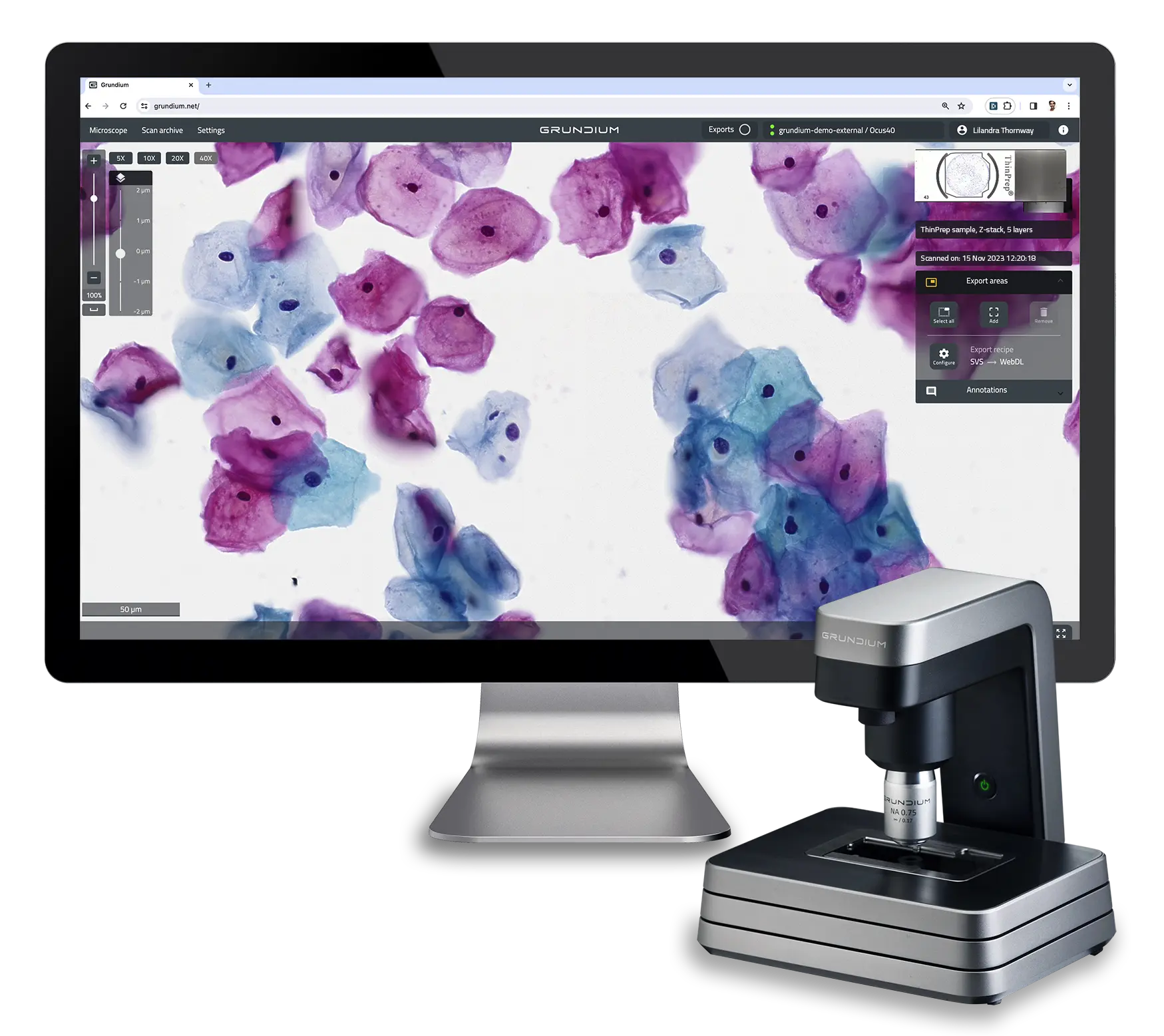
Digital pathology systems are designed with adaptability in mind, ensuring seamless integration into various workflows and laboratory environments. Modern digital pathology solutions, such as Grundium’s Ocus single-slide scanners, can often be integrated with laboratory information systems (LIS) and electronic health records (EHR), enabling efficient data exchange and reducing manual data entry. The flexible software solutions can be customized to fit specific laboratory needs, enhancing the overall efficiency of diagnostic workflows. This adaptability ensures that digital pathology systems can meet the diverse requirements of different pathology departments, from small clinics to large hospital laboratories. Collaborative efforts between IT departments, vendors, and pathology teams are essential for successful implementation and optimization.

Future Research and Development

Ongoing research and development in digital pathology focus on improving imaging technologies, developing advanced AI algorithms, and exploring new applications. Collaborative research initiatives between academic institutions, healthcare organizations, and technology companies drive innovation and accelerate the adoption of digital pathology solutions. Staying informed about the latest developments and participating in research projects can help organizations remain at the forefront of digital pathology advancements.

By addressing these considerations and adopting a strategic approach, healthcare organizations can maximize the benefits of digital pathology, improving diagnostic accuracy, efficiency, and patient care. As digital pathology continues to evolve, its integration into clinical practice will undoubtedly transform the landscape of medical diagnostics.

About Grundium

Grundium is transforming digital microscopy with our cutting-edge Ocus® whole slide scanners. Leveraging decades of expertise in high-performance imaging solutions, we deliver unparalleled image quality, user-friendly operation, and affordability. Ideal for pathology, laboratory sciences, research, and education, our scanners reflect patented technology that offers high-performance imaging at significantly lower costs.

Discover the Grundium difference, where decades of innovation and excellence elevate the standard of microscopic examinations with precision and reliability​​​​.