
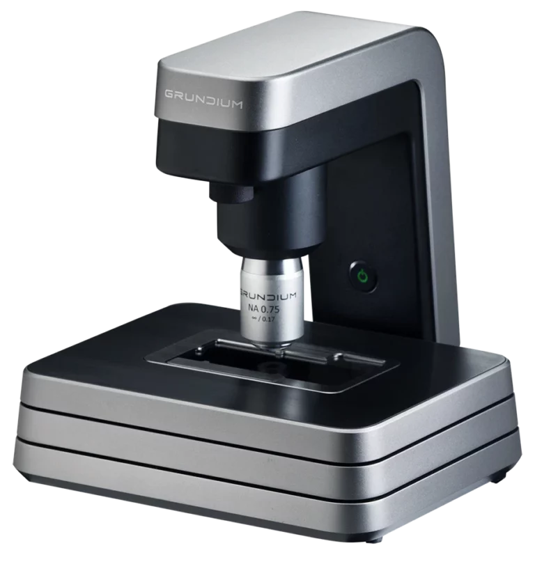
The Grundium Ocus® 20 sets new standards in digital pathology scanning, designed for speed and efficiency in histology applications.
With a focus on scan speed over higher magnification, the Ocus® 20 is ideal for intraoperative frozen section procedures, allowing pathologists to make quick, reliable diagnoses.
Combining high precision with ease of use, the Ocus® 20 offers an optimal blend of quality and affordability for labs where higher magnification isn’t necessary.
The Ocus® 40, with its powerful 40x magnification, delivers unmatched detail in every scan, making it ideal for cytology and other applications that require high-resolution imaging. For those who need the best image quality, the Ocus® 40 brings the microscopic world into sharp focus.
Engineered for accessibility and performance, the Ocus® 40 combines quality, affordability, and user-friendliness, unlocking new possibilities in digital pathology.
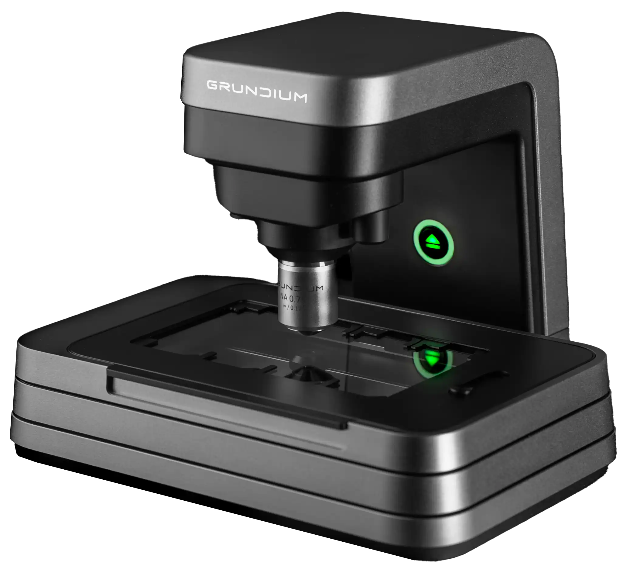
The Ocus® M series devices are Grundium’s most advanced digital pathology scanners, built for high-performance labs. With a larger slide capacity for extended walkaway scanning, and a removable slide tray for easy handling, it streamlines workflows and reduces operator time.
The intuitive, feature-rich interface makes the Ocus® M ideal for cytology and other high-magnification applications, delivering outstanding image quality and flexibility for digital pathology.
The Ocus M comes in two different magnification variants; Ocus M 20 and Ocus M 40.


Out of all the scanners I’ve used — and I’ve used a lot — it’s the easiest one. We've scanned more than 30 000 slides without any technical issues.

With Grundium Ocus-enabled telepathology, we can provide better service to our customers; the hospitals. For the hospitals the process doesn’t change, but they benefit from Fimlab working faster and more accurately with digital pathology.

Not only has Grundium created something unique in the market with the beautifully designed small-footprint scanner, but they also have a super technical team that can work really close together to make the whole solution seamless and is willing to invest themselves in making something new. It’s super unique in the world to have everything from the region of interest, focus layers, depth of field and the whole experience totally integrated.

The Ocus scanner is small and produces very sharp images fast. It is a vital instrument in field use in organ procurement. Its unparalleled portability helps save lives.

We have excellent images with the Ocus and I couldn’t be happier to have this little helper on my desk here. I use it mainly as a microscope, to be honest, but of course, scan my slides as well. It has really helped me so much.

The Philips IntelliSite Digital Pathology Solution has the capabilities to upload, view and annotate the images produced from the Grundium scanner. Zooming and panning is really fast, even when the images are physically stored on a server in the Netherlands and I’m viewing the images from a workstation in Sweden.
Discover how Grundium’s digital pathology scanners can enhance your workflow.
Request a demo or send us a message below.
When selecting a microscope slide scanner for pathology workflows, important considerations include image quality, magnification, scanning speed, workflow compatibility, slide capacity, supported file formats, and ease of use. The Grundium Ocus® series provides flexible, browser-based digital pathology solutions ranging from compact single-slide systems to four-slide Ocus® M Series scanners for workflows that benefit from additional walk-away capacity.
Yes. The Grundium Ocus® series supports whole slide imaging by digitizing microscope slides into high-resolution digital images that can be viewed, navigated, exported, and shared. This supports digital pathology workflows across applicable clinical, research, education, and collaborative review environments.
Ocus® digital pathology scanners enable digitized slides to be viewed, navigated, and shared remotely through a web browser without additional viewing software. This supports remote consultations, second opinions, multi-site collaboration, and flexible pathology workflows while helping reduce the need to transport physical slides between locations.
The Ocus®20 and Ocus®40 are compact single-slide digital pathology scanners designed for efficient and accessible slide digitization. The Ocus®20 offers 20x magnification and fast scanning for histology-oriented workflows, including frozen section and intraoperative consultation use cases where speed is important. The Ocus®40 offers 40x magnification for workflows that require higher-resolution imaging, including cytology and detailed cellular review.
The Ocus® M Series adds four-slide capacity and a removable slide tray for laboratories that need additional walk-away scanning capacity or routinely handle multiple slides per case. It maintains the compact, browser-based design philosophy of the Ocus® platform while adding multi-slide workflow features. The Ocus M series is available in both 20x and 40x magnification.
The Ocus®40 and Ocus® M 40 are designed for workflows that require high-resolution digital imaging and detailed cellular visualization. With 40x magnification and 0.25 µm/pixel scan resolution, they are well suited for cytology and other applications where fine detail is important for review, collaboration, and digital pathology workflows.
The Ocus®20 is designed for fast and efficient histology workflows where rapid access to digital slides is important. Its 20x magnification, 0.50 µm/pixel scan resolution, and approximately one-minute scan time for a 15 × 15 mm area make it well suited for histology-oriented applications such as frozen sections and intraoperative consultation workflows where higher magnification is not always required.


