Improve your intraoperative frozen section workflow

Are you tired of wasting your valuable time traveling between locations or waiting near the operating room for the samples? Would you prefer to be able to get a second opinion from your colleague in challenging cases even intraoperatively?

Have an Ocus®20 scanner in a lab near the operating room, and view the frozen section samples on your screen within minutes – and ask your colleague to participate, wherever they are. Utilizing Ocus® digital scanners saves your time for the most important part of your work and makes patient care faster and more precise.
Grundium Ocus®40 microscope slide scanner

OCUS®20 IS designed for frozen section procedures

The Ocus®20 whole slide scanner is a pivotal advancement in digital pathology, specifically designed to enhance frozen section workflows. This innovative tool combines speed, accuracy, and ease of use, making it an essential asset for pathologists and lab technicians dealing with the critical nature of intraoperative consultations.

High-QUALITY IMAGES

The Ocus®20 delivers exceptional image quality with 20x magnification and advanced optics, ensuring detailed, sharp scans for precise tissue analysis. It produces crystal clear images, enhancing diagnostics with consistently high-resolution outputs for confident observation and analysis.

EASE OF USE

The Ocus®20 offers unparalleled simplicity with its browser-based UI for direct scanning and slide management, compact design for easy lab integration, remote use for global collaboration, and flexible export options, streamlining digital pathology operations efficiently.

COST-EFFECTIVE

The Ocus®20 makes digital pathology accessible at a fraction of traditional costs. Its minimal maintenance, quick setup, and efficient operation reduce both initial and ongoing expenses, offering an economical solution for labs aiming to modernize or expand their digital capabilities efficiently.

High-quality images from frozen section slides

The Ocus®20 features fast electronic autofocus to ensure detailed focus across varied sample surfaces, crucial for the nuanced demands of frozen sections. This capability, combined with unique stitching techniques, produces seamless whole slide images without stitching artifacts, enhancing diagnostic accuracy.

High-quality optics enable the scanner to excel with a wide range of sample types, accommodating even partially dry slides thanks to its open stage design. Advanced imaging technology further ensures sharp, detailed captures at high magnification, providing pathologists with the clarity needed for precise tissue analysis.

Together, these features make the Ocus®20 a powerful ally in modern pathology, optimizing both the speed and accuracy of frozen section diagnostics.
Ocus®20 intraoperative frozen section skin sample
Skin tissue sample scanned with the Ocus20

Compact design

The compact footprint of the Ocus scanners is a key feature that sets them apart in the field of digital microscopy. By occupying minimal desk or lab space, these scanners offer a significant advantage, especially in environments where space is at a premium. This small footprint ensures that even the most cluttered laboratories or tight workspaces can accommodate an Ocus scanner without the need to reorganize or sacrifice other essential equipment.

The open-construction design of the Ocus scanner not only enhances its aesthetic appeal and makes it incredibly easy to clean the stage. If you're concerned about space, you'll be pleased with Ocus®20's compact design, making it a perfect fit even in the most cluttered labs.
The Ocus® dimensions

Remote use enables easy collaboration

The Ocus®20's capability for remote use significantly enhances collaborative efforts among medical professionals, particularly pathologists. This feature allows for the real-time sharing and analysis of high-quality digital slides, making it possible for experts to collaborate on diagnoses from different locations. Whether it's a complex case that benefits from a second opinion or a routine analysis that requires confirmation, the Ocus®20 facilitates a seamless exchange of insights and expertise.
Moreover, this remote capability is not just about sharing images; it's about fostering a more integrated approach to pathology. By enabling easy access to digital slides, the Ocus®20 breaks down geographical barriers, allowing specialists to contribute their knowledge and skills regardless of their physical location. This level of collaboration can lead to more accurate diagnoses, enhanced learning opportunities, and ultimately, better patient care. The Ocus®20, therefore, is not just a tool for digitizing slides; it's a platform for collaborative excellence in the field of pathology.
Intraoperative frozen section workflow with Ocus® scanners

Integrability to existing pathology workflows

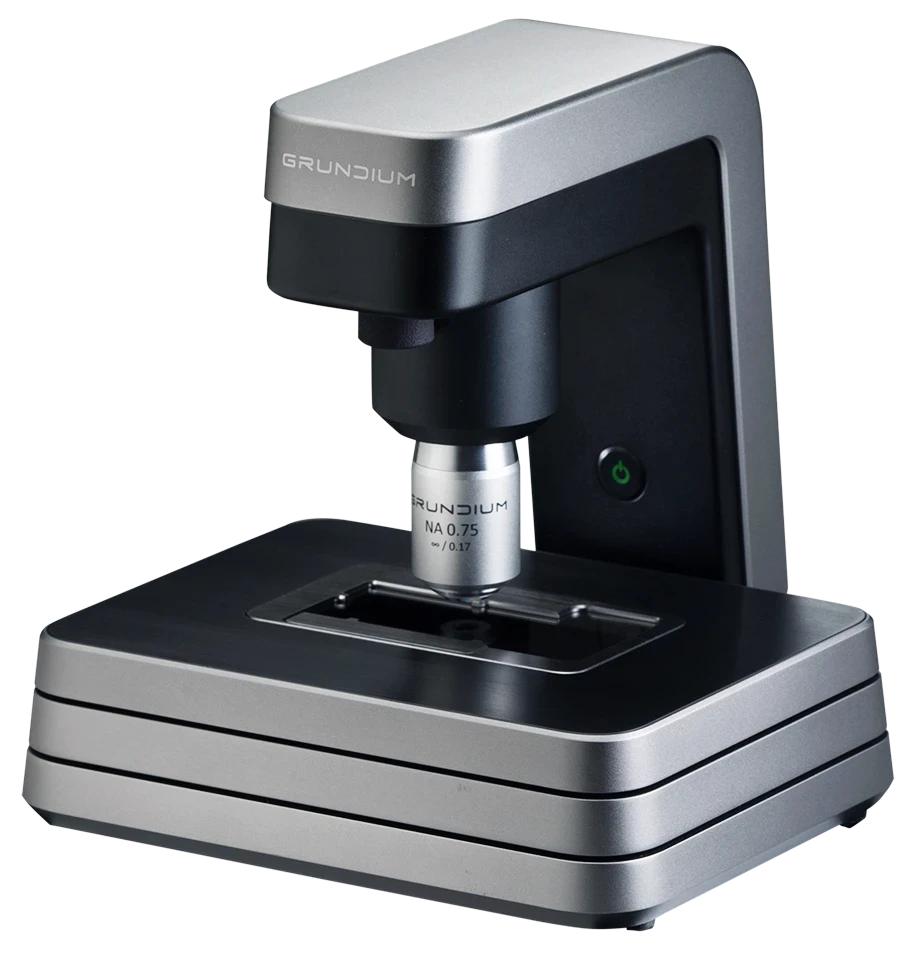
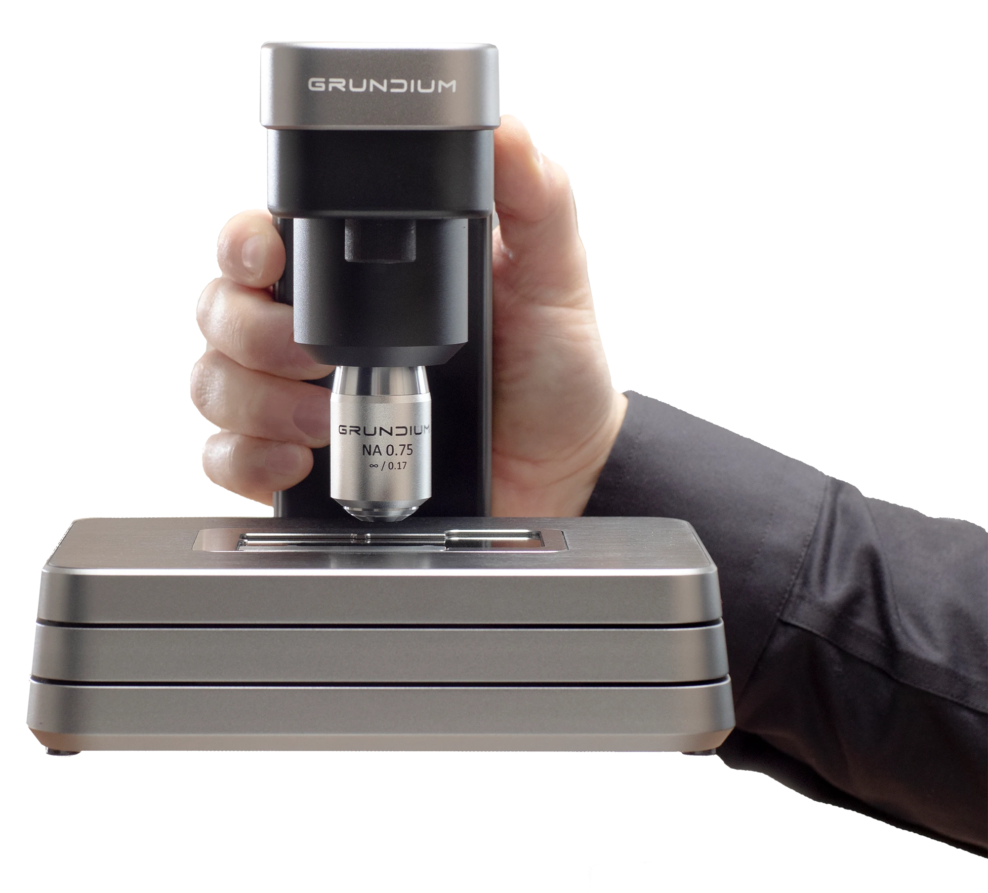
The Ocus®20 integrates seamlessly into existing workflows, thanks to its compact size, durable construction, and streamlined user interface. Its design enables rapid setup, taking just 15 minutes from unboxing to full operation, minimizing disruption to ongoing lab activities. This ease of use extends to its operational capabilities, where the intuitive, browser-based interface allows for hassle-free scanning and image sharing without the need for additional software installations.

Furthermore, the Ocus®20 supports multiple image export options and is compatible with widely used whole slide image (WSI) formats, simplifying the integration into existing digital pathology ecosystems. Its secure remote connectivity feature enhances collaborative diagnostics by enabling users to control the scanner, view images, and collaborate with experts from any location, further integrating the scanner into telepathology networks.

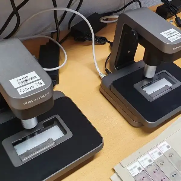
Designed with minimal maintenance requirements and a small footprint, the Ocus®20 does not just fit into physical spaces within labs easily but also adapts to the operational flow, ensuring that labs can maximize their efficiency without the need for extensive reconfiguration of their existing setup. This adaptability, combined with the scanner's cost-effectiveness and high-quality imaging capabilities, makes it an ideal choice for labs looking to transition into digital pathology or expand their current capabilities with minimal overhead.

Fimlab validated a remote intraoperative frozen section process

Fimlab is one of the leading laboratory service providers in Finland. Serving an area with 1.3 million people, Fimlab runs over 15 million diagnostic tests per year in their 120 locations.

To maximize the time the pathologist spends on what they’re indispensable at — diagnosing — Fimlab has successfully piloted a digital frozen section process operated by laboratory technicians.

Want to see the Ocus® scanners in action?

We'll gladly show you in an online demo!
A Grundium expert will arrange a 30-minute session tailored to your schedule. During the demo, we'll discuss the best Ocus® scanner for your needs, introduce you to our pricing, share user stories, and guide you on enhancing your lab's digital pathology journey.