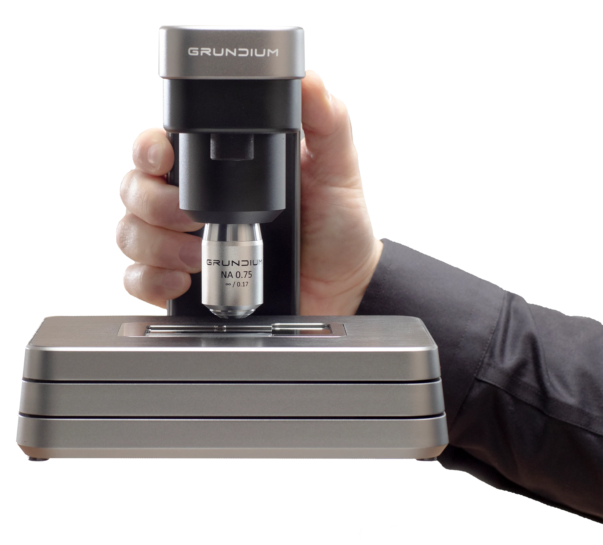
A recap of CAP TODAY’s webinar with The Ohio State University Wexner Medical Center on how laboratories can begin digital pathology adoption with focused, practical workflows. Topics include frozen sections, validation, staffing, 20x vs 40x scanning, turnaround time gains, and building a foundation for AI.
Digital pathology adoption does not have to begin with a massive, all-at-once rollout. For many laboratories, the more practical path is to start with one clear workflow problem, introduce digital tools where they fit naturally, and expand from there.
That was the central message of the CAP TODAY webinar, Simplifying Digital Pathology: Getting Started Without a Complex Rollout, broadcast on August 26, 2026. The session featured Scott Hammond, Sandra Banky, Ursula Hofer, and Molly Epler from The Ohio State University Wexner Medical Center Department of Pathology, with Bob McGonnagle of CAP TODAY moderating.
Skip recap and jump directly to the webinar recording
One of the strongest themes from the webinar was that digital pathology adoption should begin with workflow need. The OSU team emphasized that their goal was never to be “digital for digital’s sake.” The goal was to address practical constraints such as limited space, pathologists spread across multiple locations, and turnaround time pressure.
That framing is important for any lab considering digital pathology. A complex rollout can feel intimidating, especially when teams are already managing staffing, space, IT, and operational pressures. But a focused first use case can make adoption more manageable.
The best starting point is a workflow where the need is clear, the scope is contained, and the benefit is easy for staff and pathologists to understand.
For OSU, frozen sections in the gross room offered a practical starting point. Frozen sections are familiar to many hospitals, they are time-sensitive, and they often require fast access to a pathologist.
Instead of redesigning the lab around the scanner, the team incorporated scanning into the existing process. A specimen still comes from the OR. The case is accessioned. The slide is prepared. Then, instead of physically handing the slide to a pathologist in the same location, the slide can be scanned and reviewed remotely.
That distinction matters. Digital pathology becomes easier to adopt when the technology fits into the existing workflow rather than forcing the workflow to bend around the technology.
The webinar made clear that successful digital pathology implementation is not just a technical project. It is a change-management project.
Ursula Hofer described adoption through four pillars: training, standardization, validation, and building confidence. Staff and pathologists need to know how to operate the system, but they also need to understand how it fits into their daily work.
Molly Epler reinforced the same point from an operations perspective. Training should be practical and role-specific. Teams do not need to be overwhelmed with every feature on day one. They need to know what they will actually use in their routine workflow.
Simple support systems can make a difference: clear policies, one-page cheat sheets, internal super users, and a plan for answering questions when staff encounter friction. As the OSU team noted, questions and pain points are a normal part of change. They do not mean the technology has failed.
Validation was another major theme. Before a scanner is used clinically, laboratories need confidence that the technology performs as expected and supports patient care.
The OSU team described a structured validation process: selecting representative cases, de-identifying slides, scanning them on the intended device, comparing glass and digital review, adjudicating discrepancies, and maintaining complete documentation.
This kind of validation is not just a compliance exercise. It helps turn a new workflow into a clinically accepted process. It also gives pathologists and laboratory teams confidence that digital review can support the work they need to do.
One of the most compelling parts of the webinar was the discussion of access. Digital pathology can help bring subspecialty expertise to the right case at the right time, even when pathologists are not in the same building, hospital, or city.
The OSU team shared examples from community and regional workflows. In one Q&A exchange, Scott Hammond noted that a community site saw turnaround time improve by about 23 hours after adopting digital pathology. Sandra Banky added another striking example: a digital consult that might normally have taken two to three days using glass slide transport was completed in about five minutes.
For labs evaluating digital pathology, those examples are powerful because they connect the technology to an operational and clinical outcome: faster access to the expertise needed for timely decision-making.
The Q&A also explored a practical question many laboratories face: should teams use 20x or 40x scanning?
The answer depends on the workflow. For frozen sections, speed often matters most. Josh Griffith of Grundium noted that, using a standard benchmark, a 15 by 15 millimeter tissue section takes about one minute to digitize at 20x and about three to three and a half minutes at 40x.
The OSU team described a similar practical tradeoff. For some frozen section workflows, 20x may provide the speed needed to keep turnaround time tight. For other situations, pathologists may prefer 40x because it offers more detail and confidence. In some cases, live view may be the right option.
The broader lesson is not that one magnification is always right. The lesson is that scanner settings should be chosen based on the clinical workflow, time pressure, pathologist preference, and diagnostic need.
Staffing was another audience question, and the answer was appropriately nuanced.
For frozen sections and rapid on-site evaluations, OSU did not need to hire additional technologists specifically because telepathology was incorporated into the existing workflow. However, broader prospective scanning is different. Scott Hammond described dedicated scan techs who support clinical sign-out workflows, and Sandra Banky described additional operational support needed to manage slides, scanning, distribution, filing, and pathologist preferences.
That nuance is important. A focused digital pathology starting point may fit within current staffing. A broader digital operation may require a more formal staffing and support model.
The webinar also addressed the question of what “fully digital” means. Scott Hammond defined it in practical terms: having a pipeline that allows a pathologist to sign out a case clinically in some capacity.
That does not necessarily mean every case, every division, and every site becomes digital at the same time. At OSU, different divisions and locations introduced digital workflows at different points, depending on operational needs and infrastructure readiness.
IT support is essential in that process. Network capacity, image management, remote access, and system performance can all affect implementation. Digital pathology is a clinical workflow, but it is also an infrastructure workflow.
The webinar closed the loop between practical adoption today and the future of pathology. Computational pathology and AI depend on digital slides and robust digital workflows. Labs do not need to reach that future overnight, but they do need a path toward it.
Starting with a focused workflow can create that foundation. A lab might begin with frozen sections, cytology, consults, or another manageable use case. Over time, early success builds confidence, reveals additional opportunities, and prepares the organization for more advanced digital and computational tools.
A good first use case is a focused workflow with a clear operational need. Frozen sections and rapid on-site evaluations are strong examples because they are time-sensitive, familiar, and benefit from remote access to pathologists.
No. The OSU team emphasized that labs can start small. The key is to identify one workflow problem, choose technology that fits that workflow, train users, validate the process, and expand from demonstrated success.
It depends on the scope. OSU did not need additional staff specifically for frozen section telepathology or rapid on-site evaluations, but broader prospective scanning required dedicated scan techs and operational support.
The choice depends on the use case. For frozen sections, 20x is often preferred because it is faster. For workflows that require additional detail or pathologist confidence, 40x or live view may be more appropriate.
AI and computational pathology depend on digitized slides and reliable digital workflows. A focused digital pathology implementation can help laboratories build the foundation needed for future AI-enabled workflows.
Digital pathology adoption does not have to be intimidating. The most practical starting point is often not the biggest transformation, but the clearest problem.
For laboratories exploring how to begin, the OSU experience offers a useful model: start with a focused workflow, make the technology fit the process, support the people using it, validate carefully, and build toward the future one successful use case at a time.
This AI-assisted recap was prepared from the webinar and reviewed by the Grundium team before publication.